 If there was one experience that pushed my interest in neurobiology beyond the activation threshold and kick-started the process that led to the creation of this blog, it was reading Dr. R. Douglas Fields' article "Making Memories Stick" in the Feb. 2005 issue of Scientific American (ref.). I'd long been interested in molecular biology but had been intimidated by the level of jargon and assumed knowledge that filled most articles. Dr. Fields' article explained the inner workings of a neuron so clearly and lucidly that I was able to get a basic understanding of what was happening, and was motivated to try to learn more about molecular biology and neurobiology in particular. This blog is essentially the notes I've been making as I try to learn more about the details of how biology works at a molecular, cellular and neuronal level. So, things have finally come full circle. Let's take a deeper look at what makes memories stick...
From Making Memories Stick by R. Douglas Fields: Remarkably, if the same high-frequency stimulus is applied repeatedly (three times in our experiments), the synapse becomes strengthened permanently, a state called late LTP. But the stimuli cannot be repeated one after the other. Instead, each stimulus burst must be spaced by sufficient intervals of inactivity (10 minutes in our experiments). And adding chemicals that block mRNA or protein sysnthesis to the salt solution bathing the brain slice will cause the synapse to weaken to its original strength within two to three hours. Just as in whole organisms, the cellular model of short-term memory is not dependent on the nucleus, but the long-term formation of memory is.
As far back as 1949, a psychologist named Donald Hebb ...proposed that, like an orchestra player who cannot keep up, a synapse on a neuron that fires out of sync with the other inputs to the neuron will stand out as odd and should be eliminated, but synapses that fire together - enough so as to make the nueron fire an action potential - should be strengthened. The brain would thus wire itself up in accordance with the flow of impulses through developing neural circuits, refining the original general outline.
...
By stimulating neurons to fire action potentials in different patterns and then measuring the amount of mRNA from genes known to be important in forming neural circuits or in adapting to the environment, we found ...we could turn on or off particular genes simply by dialing up the correct stimulus frequency on our electrophysiological stimulator, just as one tunes into a particular radio station by selecting the correct signal frequency.
...
If [the hippocampus] is dissected from a rat and kept alive in a salt solution, microelectrodes and electronic amplifiers can record the electrical impulses from individual synaptic connections on a neuron. By administering a burst of electrical shocks to a synapse, causing it to fire in a specific pattern, that synaptic connection can be strengthened. That is to say, the synapse produces about twice as much voltage in response to subsequent stimulations after it has received the high-frequency stimulus.
This increased strength, termed long-term potentiation (LTP), can be, despite its name, relatively short-lived. When test pulses are applied at a series of intervals after the high frequency stimulus, the voltage produced by the synapse slowly diminishes back to its original strength within a few hours. Known as early LTP, this temporary synaptic strengthening is a cellualr model of short-term memory.
...
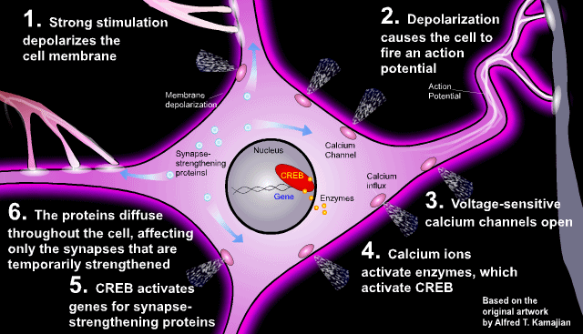
Strong stimulation, either from the repeated firing of a single synapse or from the simultaneous firing of several synpases on a cell, depolarizes the cell membrane, causing the cell to fire action potentials of its own, which in turn causes voltage-sensitive calcium channels to open. The calcium ions interact with enzymes that activate the transcription factor CREB, which activates the genes for manufacturing synapse-strengthening proteins. The cell's nucleus "listens," in effect, to the cell's output - firing action potientials - to determine when to permanently strengthen a synapse and make a memory last.
More info on the role CREB plays is available here.)
...
One does not always know beforehand what events should be commmited permanently to memory. The moment-to-moment memories necessary for operating in the present are handled well by transient adjustments in the strength of individual synapses. But when an event is important enough or is repeated enough, synapses fire to make the neuron in turn fire neural impulses repeatedly and strongly, declaring "this is an event that should be recorded". The relevant genes turn on, and the synapses that are holding the short-term memory when the synapse strengthening proteins find them, become, in effect, tattooed.
(Some recent research that explains how this 'tattooing' works will be covered in the next post). From another excellent article by Dr. Fields, Erasing Memories (SciAm Mind Dec 2005):
There is a complication, however. The molecules that establish current flow around synapses are proteins, and all proteins in the body degrade and are replaced constantly over a period of hours or days. To strengthen a neural connection for a lifetime, some other process must take place to bolster the physical structure of a synapse or form additional synapses between the neurons involved.
The transition from temporary to permanent memory is called consolidation. Many experiments have deterined that consolidation requires many hours, and it can be enhanced or blocked in various ways. ... If the next 10 seconds of your life could be your last, you will remember them, even if the interval is folowed by another 10 dramatic seconds, and so on. ... The heightened state of attention, stress and novelty stimulates the consolidation phase of memory.
Neuroscientists have discovered how this consolidation happens. An epinephrine (a.k.a. adrenaline) rush releases a flood of stress hormones and neurotransmitters that activate the amygdala, the brain region that processes fear and emotion. The amygdala connects to many other regions where different kinds of memories are stored, and it boosts incoming data that have emotional impact. Consolidation, therefore, possibly can be aided by increasing levels of these neurotransmitters or hormones.
...
A growing body of evidence suggests that memory consolidation continues "off-line" while we sleep, in part because sleep involves periodic surges of some of the same hormones and neurotransmitters that are aroused in stressful and novel situations.
A research group headed by Jan Born at the University of Lübeck has found that sleep not only strengthens the content of a memory, but also the temporal structure of episodic memories, probably by replaying them in the forward direction (ScienceDaily (Apr. 18, 2007))
Returning to Erasing Memories:... [Work by Phillipe Peigneux] illustrates that memory consolidation requires sorting through fresh memories, integrating them with other memories, and shuttling them to different brain regions for permanent storage. Short-term memories deemed dispensible are discarded.
...
[James R. Misanin and others at Rutgers University] found that a consolidated memory could be erased if a lab rat was shocked right after being forced to recall the experience. ... [R]ecalling the memory had somehow made it vulnerable to disruption. This phenomenon has been termed reconsolidation.
In the article, Dr. Fields mentions that research by Joseph E. LeDoux and his colleagues at New York University has shown that microinjecting protein synthesis inhibitors into a rat's amygdala can block memory reconsolidation if applied shortly after the memory is recalled.
From Joseph LeDoux's book The Synaptic Self: The recent discovery , made by Karim Nader and Glenn Schafe in my lab, is that protein synthesis in the amygdala seems necessary for a recently activated memory to be kept as a memory. That is, if you take a memory out of storage you have to make new proteins (you have to restore, or reconsolidate it) in order for the memory to remain a memory. One way of thinking about this is that the brain that does the remembering is not the brain that formed the initial memory. In order for the old memory to make sense in the current brain, the memory has to be updated.
From Cellular and Systems Reconsolidation in the Hippocampus by Jacek Debiec, Joseph E. LeDoux and Karim Nader:
Indeed, reconsolidation and consolidation have been found to share a number of common properties, including: (1) requirement of protein synthesis in order for the memory to persist, (2) time windows during which protein synthesis blockade is effective, and (3) that protein synthesis blockage in the same brain region, the amygdala, disrupts both. Given these similarities, it seemed parsimonious to conclude that a new memory and a reactivated, consolidated memory share a common memory state, as originally proposed by Lewis (1979). Thus, instead of just occurring once, memory storage may instead be a process that is reiterated with each use of the memory.
... and from the abstract:
Cellular theories of memory consolidation posit that new memories require new protein synthesis in order to be stored. Systems consolidation theories posit that the hippocampus has a time-limited role in memory storage, after which the memory is independent of the hippocampus. Here, we show that intra-hippocampal infusions of the protein synthesis inhibitor anisomycin caused amnesia for a consolidated hippocampal-dependent contextual fear memory, but only if the memory was reactivated prior to infusion. The effect occurred even if reactivation was delayed for 45 days after training, a time when contextual memory is independent of the hippocampus. Indeed, reactivation of a hippocampus-independent memory caused the trace to again become hippocampus dependent, but only for 2 days rather than for weeks. Thus, hippocampal memories can undergo reconsolidation at both the cellular and systems levels.
Do you remember this quote, near the start of Dr. Fields' article 'Making Memories Stick': Donald Hebb ...proposed that, like an orchestra player who cannot keep up, a synapse on a neuron that fires out of sync with the other inputs to the neuron will stand out as odd and should be eliminated.?
The official term for the weakening of unhelpful synapses such as these is Long Term Depression (LTD). From the Howard Hughes Medical Institute (2004) LTP and LTD: strengthening and weakening synaptic connections
OUT OF SYNC, LOSE THE LINK
"The prevailing view used to be 'use it or lose it,'" says Bear. But a more accurate conclusion may be "neurons that fire out of sync lose their link.
...
When a neuron receives mismatched signals, synapses lose receptors ... If the loss of receptors is sufficiently prolonged, Bear suspects, the synapse eventually will disappear.
...
Bear's research into LTD has had other, and sometimes quite unexpected, consequences. His lab also has investigated a form of LTD triggered by the activation of another receptor on receiving neurons. One of the proteins affected by this receptor is the one that's missing in fragile X syndrome, the most common cause of inherited mental retardation in humans.
As always, the more I learn about things the more questions I have! Next post, we'll zoom in further to look at the changes the synapse undergoes when it is 'strengthened', and then start to zoom out to look at memory from a higher 'system-level' perspective.
More...
Podcast interview with R. Douglas Fields
Synapse signalling complexes and networks: machines underlying cognition" by Seth G.N. Grant
Comments